Image-guided irradiation planning software

Features

Intuitive, powerful,
designed for research

Use all capabilities of your image-guided radiation research platform for complex radiobiological targeting; plan, quantify, evaluate and report with the most reliable radiation calculation methods.

clinical_notes

Research-minded

A small learning curve, visual workflow, quick export of dose-volume metrics, save any shown irradiation plan visually to a high-resolution image, ready to be included in your report or manuscript

finance

Easy beam planning

Add beams and irradiation targets in an interactive manner, click-and-drag to quickly create an irradiation plan, immediately find out how irradiation beams will traverse the 3D image and all volumes of interest

list_alt_check

Reliable irradiation calculation

Using high-resolution CTs and proper calibration and validation to ensure accurate irradiation quantification. The DICOM standard can be used to link inputs and outputs, ideal for longer term archival of research data

edit_road

Built-in segmentation

Machine-specific calibrations and built-in 3D segmentation tools to define all relevant parts of the irradiation volume, defined manually or using our AI-powered automatic segmentation tools

FAQ

Frequently
asked questions

01.

Can I use our other preclinical imaging  modalities to enhance radiation planning  and evaluation?

keyboard_arrow_down

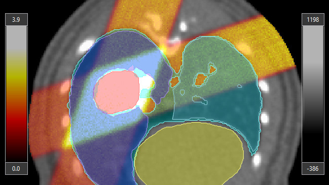
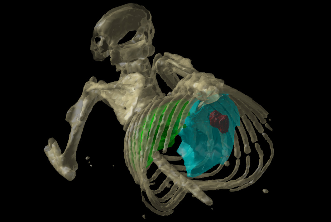
Yes. Multiple preclinical CT, MR, PET, Optical Tomography, and SPECT formats can be imported as secondary 3D image data volumes. While a primary planning CT image is mandatory, secondary image data volumes can be manually registered to the primary CT image and used during segmentation and planning to define points and volumes of interest.

02.

Is SmART-TPS compatible with my image-guided irradiation research platform?

keyboard_arrow_down

Probably. Our suite of applications is tested and validated with most versions of the image-guided irradiation platforms of the major international vendors. For custom or one-off platforms, we can assess or develop compatibility if the platform allows for the required data sharing capabilities of inputs and outputs. For further inquiries, don't hesitate to contact us.

03.

How does the AI-based autocontouring module work together with SmART-TPS?

keyboard_arrow_down

Our autocontouring module, SmART-Contour, is a fully integrated module of the SmART-TPS irradiation planning software. Segmentation can start automatically in the background once a CT is loaded, and created volumes of interest can be used for irradiation planning and evaluation once the user accepts the structures.

04.

Can we use SmART-TPS to plan our irradiations of cell cultures, vials, and other ex-vivo specimens?

keyboard_arrow_down

Yes. Irradiation planning is CT-image based; the specimens must be included in the CT image and correct media and mass densities are required as usual. However, specific defaults can be set for such studies. If a larger irradiation field is required further from the X-ray source, model-based virtual planning is required; we anticipate supporting that in a future release.

Alternatively, our non-image-guided treatment planning software was created for this specific purpose.

arrow_outward